It was not what I expected to hear at my routine mammogram—as the radiologist talked, all I could think was, “What the…?!”
But what seemed like the worst day ever may have been my lucky break. That mammogram in my late 40s had detected a warning sign: a tiny cluster of calcification in my left breast. I needed a stereotactic core needle biopsy, a procedure where the suspect spot is located via mammogram, extracted with a special needle, and sent to a pathology lab to see if the tissue is normal or not.
Chances are you know someone like me.
More than 1 million women have breast biopsies each year in the United States, most of which are core needle biopsies (CNB), according to the Agency for Healthcare Research and Quality. If you have a suspicious finding on a breast imaging exam, don’t panic. “The overwhelming odds are you don’t have cancer,” explains Nina Vincoff, MD, chief of the division of breast imaging at Northwell Health. “At least three quarters of the time, your biopsy is going to turn out to be benign.”
Still, it’s essential to get that biopsy just in case you do have cancer or precancerous cells. So what does the procedure actually entail, from prep to recovery? Does it hurt? Can you go back to work after? Here’s exactly what to expect.
The core needle biopsy breakthrough
Just a few decades ago, if you needed a breast biopsy, it meant either surgery or a fine needle aspiration biopsy—an imperfect test where a thin needle extracts a very small sample of cells. These days, the majority of breast biopsies are the core needle kind, and that’s a good thing. “It’s the far preferred way of doing a breast biopsy because you get much more specific results than you do with a fine needle aspiration without having to go through surgery,” Vincoff says.
The name “core needle” refers to the needle itself: It’s hollow, meaning it can grab the target and surrounding tissues. And, Vincoff adds, a core needle specimen is big enough to give a lot of information—not just whether or not you have cancer, but also the specific type of cancer, plus a snapshot of the health of the surrounding breast tissue.
What the heck is a "stereotactic core needle breast biopsy” anyway?
So meanwhile, back at my routine-mammo-from-hell, my head was spinning as the radiologist told me I needed to schedule a stereotactic core needle breast biopsy.
A what?
A stereotactic core needle biopsy, I learned, is a CNB using mammography to direct the radiologist to the target. (Depending on which screening test originally found the suspicious area, a core needle biopsy may also be guided by ultrasound or MRI. Calcifications—which are calcium deposits—are usually found on mammograms, but they’re hard to see via ultrasound and MRI.)
Before scheduling the procedure, I asked how much time I’d need off from work and was surprised to hear I could go back to my desk job the next day. “Just no going to the gym after the biopsy,” the scheduler added. When I laughed and said, “I wasn’t planning on it,” she said, “You’d be surprised how many women want to work out right after!”
Biopsy day
That morning, I showered but skipped deodorant and body lotion as instructed. When my husband and I got to the radiology office, a nurse ushered me into a room and asked me whether I could be pregnant (nope!). I was joined by the radiologist—the doctor who does the biopsy—and technologist—the provider who positions the breasts and tells you when to breathe.
Next up, I had to lie face down on a big table with cutouts for my breasts to fall through (the doctor works below you). To be honest, it felt awkward to have my boobs dangling down. Fortunately, according to Vincoff, it’s more common these days to have the procedure sitting in a chair with the mammography machine in front of you.
Next, the technologist snaps a picture of the target using mammography or whatever imaging your situation requires. Then the radiologist injects a local anesthetic. You’ll feel that part a bit: I felt a little pinch and burn, just like you do at the dentist’s office when they give you a numbing agent.
You should feel a little pressure but no pain. If you do feel pain, speak up. “I always tell people don't spare my feelings,” says Vincoff. “Everybody's different and is going to need slightly different amounts of anesthesia.”
Next, the radiologist uses the core needle to extract the samples. My radiologist and technologist talked me through each step, which really helped dial down my anxiety. After that, the doctor left the room to get an X-ray of the specimen to make sure she could see calcifications—confirmation that she hit the mark.
For the final step, the doctor puts a marker in your breast. “It’s a tiny piece of metal the size of a sesame seed or smaller,” Vincoff says. Don’t worry—you can’t feel it and it won’t set off metal detectors in the airport. Why is it there then? “If you have something that needs surgery afterwards, it guides the surgeon to know what needs to be taken out,” Vincoff explains. And even if you don't need surgery, it tells the radiology team at future imaging tests that this is an area that has already been sampled.
On to healing
It was over before I knew it.
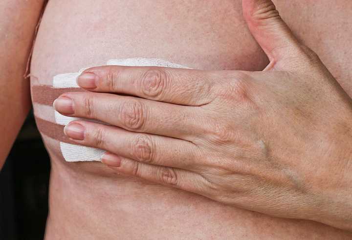
The doctor closed the spot with Steri-Strips and wrapped my breast tightly with an elastic bandage to reduce swelling. I was told to ice on and off for the first day and take Extra Strength Tylenol if needed.
For the first 24 hours, I had to keep the area dry and avoid strenuous activity. A little blood on the bandage is normal, the doctor said, but in very rare cases, complications can happen, so if I noticed significant bleeding or swelling, warmth, or hardness in the breast, I needed to go to the ER. (Thankfully that didn’t happen.)
Since my biopsy was on a Friday, I took the weekend to relax and catch up on my Netflix queue. By Monday morning, I was back at my desk, feeling normal.
The waiting is the hardest part
CNB results usually take two days to a week, depending on whether the sample needs to be analyzed further. Anywhere from 15% to 25% of the time, the results show breast tissue that will need surgery for more testing. Sometimes, the biopsy finds precancerous cells or cells that are not a pre-cancer but not completely normal, either.
In rare cases, the findings are not “concordant” with what the radiologist expected, says Vincoff. She explains that this means that “even though the results came back benign, you'll sometimes get a call from me saying, ‘I'm happy to tell you everything so far looks good, but I'm going to want you to have surgery to get a bigger sample.’”
And if you do hear that you have cancer or something in need of a closer look? Your next step will be to consult with a breast surgeon.
I know how scary that sounds, because I got that call.
The Tuesday after my test, the radiologist called: My biopsy showed DCIS, a type of stage 0 breast cancer. I was shocked, numb, terrified.
But thanks to that timely biopsy and a lumpectomy with an excellent and kind surgeon, I’m doing great three and a half years later. And I’m endlessly grateful for all the breakthroughs in breast cancer detection that meant I got diagnosed early—and got well.
